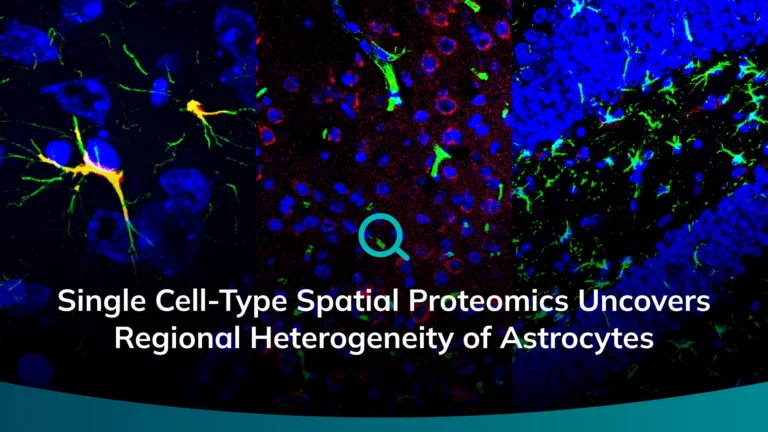
A peer-reviewed study shows Microscoop® enables microscopy-guided proteomics in brain tissue, revealing region-specific astrocyte protein signatures and candidate markers, highlighting how protein-level analysis complements transcriptomic insights.
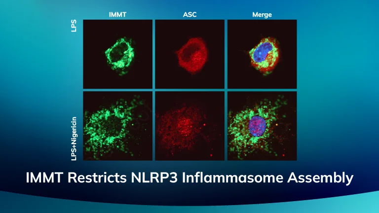
A customer publication in Molecular & Cellular Proteomics leveraged systematic interactome profiling and microscopy-guided optoproteomics to map 1,496 NLRP3- and ASC-associated protein interactions across inflammasome priming and activation states. The study identified novel inflammasome-associated proteins and highlighted IMMT as a potential regulator of NLRP3 inflammasome assembly and function
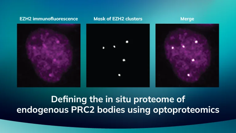
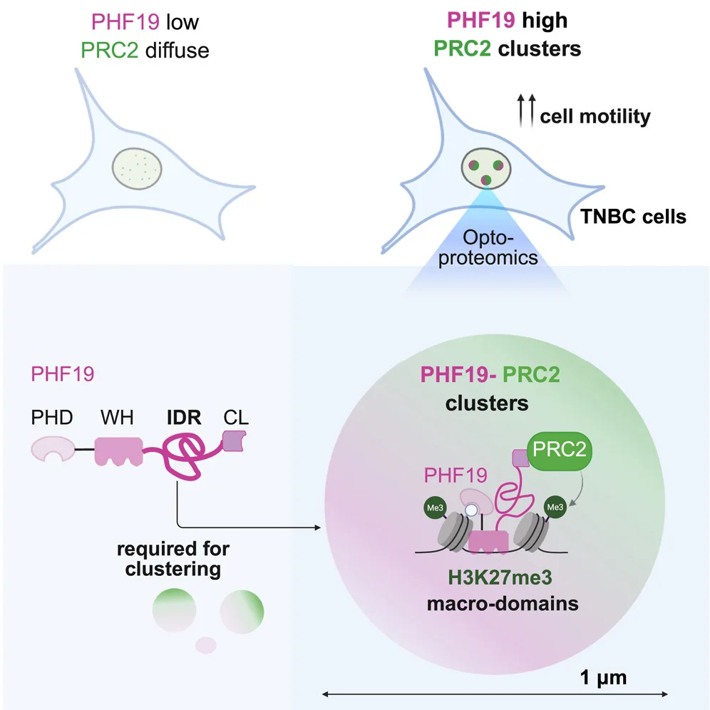
A customer publication in STAR Protocols describing a step-by-step image-guided optoproteomics workflow to profile endogenous EZH2/PRC2 bodies in breast cancer cells and map the proteomic composition of Polycomb bodies in an unbiased manner. This work showcases the potential of spatially resolved proteomics to study chromatin-associated domains and advance our understanding of chromatin organization.
This preprint identifies region-specific astrocyte proteomic signatures in the mouse cerebral cortex and hippocampus, revealing novel markers such as MINK1 and PLEKHB1 and showing how protein-level diversity supports astrocyte functional specialization.
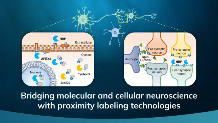
The Nature journal review (Nat Commun 2025) notes that while BAR allows profiling of endogenous proteins without genetic modification, its reliance on antibodies, broad 200–300 nm labeling radius, and use of hydrogen peroxide limit spatial precision and live-cell applications.
Microscoop Mint addresses these challenges by combining high-resolution imaging with photoactivatable biotin probes, enabling submicron, light-directed labeling. This approach improves spatial specificity and minimizes off-target background, offering researchers, professors, and PIs a practical solution for mapping protein environments in complex or fixed tissue samples.
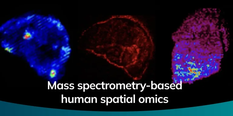
Mass spectrometry-based spatial omics integrates the molecular depth of MS with spatially resolved imaging to map proteins, metabolites, and other biomolecules directly in tissue and cells, preserving their native localization. A recent review in Journal of Biomedical Science outlines the rapid evolution of this field, from label-free mass spectrometry imaging to advanced labeling and proximity-based methods. Among these innovations, optoproteomics (Syncell’s Microscoop technology) stands out as a powerful new approach that eliminates the need for predefined targets by using image-guided photochemistry to label proteins within user-defined regions of interest, enabling unbiased, high-resolution proteomic profiling even in fixed tissue specimens.
These advances are expanding the utility of spatial proteomics across applications such as tumor microenvironment characterization, mechanistic studies of disease, and biomarker discovery — areas where retaining spatial context is critical to understanding biology. Optoproteomics and related MS-based spatial technologies are poised to accelerate discovery in oncology, immunology, neuroscience, and precision medicine by revealing spatially resolved proteome features that traditional methods cannot access.