Neuroscience

DISCOVER THE UNKNOWN PROTEINS DRIVING NEURODEGENERATION

Proteomic discovery for neurodegeneration, memory, and the neuronal microenvironment

• β-amyloid plaques
• TDP-43 aggregates
• α-synuclein bodies
• FUS aggregates
• Prion plaques
• Ubiquitin-positive inclusions
• Autophagy-related aggregates

These are signatures easily observable in neurodegenerative diseases but hard to isolate, constraining discovery of the proteome and mechanisms underlying disease.

Synaptic plasticity, multicellular interactions, and regional brain signaling involve complex protein functions. Conventional protein assays are unable to systematically isolate proteomes from these subcellular and nanoscopic regions, leaving many mechanistic questions unanswered.

Asking the impossible questions:

What defines the molecular composition of individual synapses involved in neuropathologies?
What proteins can be new biomarkers for Parkinson’s diseases?
What are new druggable targets of Alzheimer’s diseases?
What proteins are involved in storing our memories?
How do layers of entorhinal cortex communicate with each other?
What proteins are involved in specific glia-neuron interactions?

Microscoop closes these gaps by enabling in situ protein identification from nanoscale regions within intact neural systems without disrupting native cellular organization.

Nanoscopic Access to the Proteome

Microscoop combines optical targeting with proximity-based protein tagging, allowing researchers to illuminate and identify proteins within defined nanometer-scale regions of interest.

  • Nanoscopic precision: Analyze proteins down to 25 nm across neurons, synapses, and organelles paired with ROI selection.
  • Unbiased protein capture: Discover the proteome within the illuminated zone, integrated seamlessly with high sensitivity mass spectrometry.
  • Preserved biology: Study native cells, FFPE tissue sections, or aggregates without disrupting the cellular environment.

By coupling optics and proteomics, proteins can be defined not just by presence, but by “where” they operate in the cell and how those local interactions change under perturbation.

Beyond Observation to Mechanism

Microscoop enables researchers to:

  • Detect early molecular changes in neurodegeneration before disease progression.
  • Differentiate region specific signatures in neuronal cells and cellular compartments.
  • Link protein localization to trafficking and functional outcomes.

NEUROSCIENCE APPLICATIONS

01

Defining the Proteome of Amyloid-B Aggregates

Microscoop enabled automated, high resolution analysis of millions of amyloid-β aggregates in differentiated neurons, directly identifying 1,499 associated proteins. Beyond known interactors, novel candidates including Lon protease and DDX3X helicase were validated in Alzheimer’s disease mouse models. By targeting aggregates in situ without genetic modification, Microscoop uncovered new components of amyloid pathology, paving the way for next-generation biomarker and therapeutic discovery.

02

Regional Proteome Diversity of Astrocytes

Astrocytes vary widely across brain regions, shaping neural signaling and metabolism in distinct ways. Using Microscoop, researchers performed targeted photolabeling of cortical and hippocampal astrocytes, uncovering region-specific protein signatures such as CST3, FBLN5, and PSD3. This unbiased proteomic profiling revealed molecular diversity within a single cell type, showing how regional differences in astrocyte composition may influence brain function and disease vulnerability.

03

Accessing Purkinje Neuron Proteomes from Archived Brain Samples

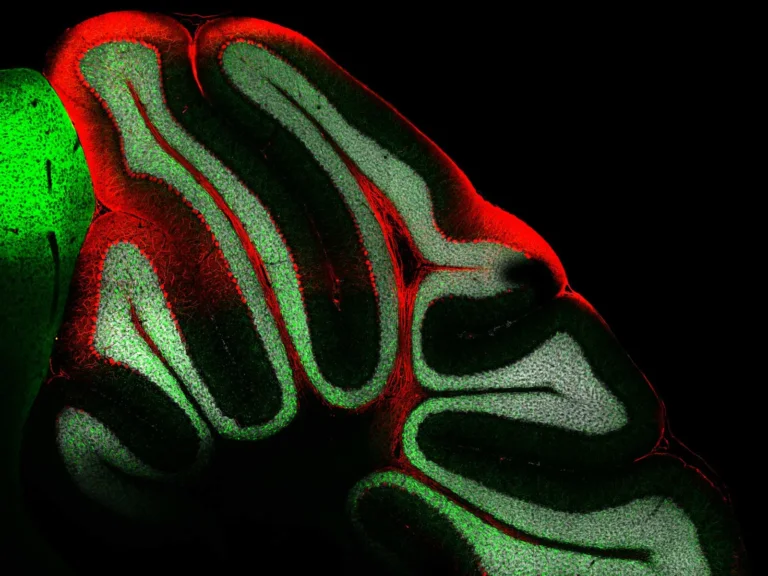
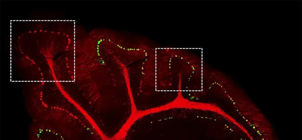
Purkinje neurons play a critical role in motor coordination and are implicated in ataxia, ALS, and other neurodegenerative disorders, yet their molecular composition is difficult to study in preserved clinical tissue. Using Microscoop, researchers performed targeted photolabeling of Purkinje cell bodies directly within FFPE mouse cerebellar sections. The workflow identified known Purkinje-specific proteins, including CALB1, GRIA1, and PKCγ, and revealed pathway enrichment in axon guidance and Rho GTPase signaling.
NEW PUBLICATION

PHF19 drives the formation of PRC2 clusters to enhance motility in TNBC cells

Pelzer, Nina et al.
Cell Reports, Volume 44, Issue 10, 116391