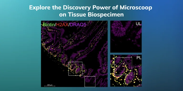
Unlocking Submicron Proteomes Spatial Proteomics Analysis with Microscoop® on Tissue Biospecimen
Microscoop® spatial proteomics enables hypothesis-free proteome identification within submicron structures in FFPE or fresh-frozen tissue sections of brain, intestine and lung tissues (among others) accelerating biomarker discovery and diagnostics and therapeutics development.
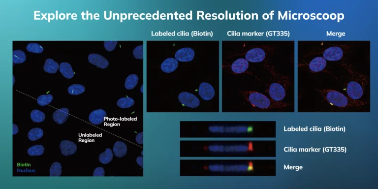
Pushing the Limits Unveiling Proteome of Primary Cilia at Unprecedented Resolution Using Microscoop®
This study highlights Microscoop®’s ability to map the proteomic landscape of primary cilia at unprecedented resolution, identifying 4,233 proteins, including 524 known ciliary proteins critical for assembly, transportation, and signaling.
Microscoop® optoproteomics (combined localization and MS) reveals spatially resolved protein complexes such as spliceosomes and histone complexes, highlighting its potential to advance subcellular biology and uncover intricate cellular interactions.
Microscoop® Two Photon-Induced Biotinylation of Protein Constituents with Submicron Specificity
Spatial proteomic discovery at specific subcellular locations often faces challenges due to limitations in current technology. Microscoop® spatial proteomics limitations by enabling hypothesis-free targeted protein identification within individual organelles, allowing exploration of subcellular protein interactions.
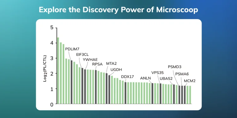
This study on stress granules (SG)s identified 1,754 consistently enriched proteins, with 74% of the top 50 ranked proteins being true positive SG proteins, underscoring Microscoop’s potential in advancing SG biology and driving therapeutic innovations.